Upper Thigh Anatomy / / Upper thigh anatomy (page 1).. There is an obvious constriction which marks the base of the head with the upper portion of the neck, and this also marks a very common site for fractures in the elderly. It occupies posteromedially in the thigh, superficial to the semimembranosus. The thigh muscles don't just move your legs. 12 photos of the muscle anatomy of upper thigh. The information contained in anatomy atlases is not a substitute for the medical care and advice of your physician.
Anyway, here r some anatomy practices for cheshire(upper thigh up(?) ). Upper part of medial surface of the shaft of tibia behind the sartorius and. There may be variations in treatment that. In human anatomy, the thigh is the area between the hip (pelvis) and the knee. It occupies posteromedially in the thigh, superficial to the semimembranosus.
Femur Definition Function Diagram Facts Britannica from cdn.britannica.com 2, vastus medialis & intermedius muscles. Muscle and tendon characteristics classic human muscles of the leg and foot classic human anatomy in motion: I'm doing some study for his body, since i want to materialize and practice perfect body for cheshire, before i start making outfits for him. The artist's guide to the. Muscle anatomy diagram front upper thigh pain symptoms lower leg muscle anatomy the hollow of thigh anatomy left hip muscle anatomy torn tendon in upper thigh parts of the leg anatomy thigh muscle anatomy cross section leg muscle anatomy model leg bones anatomy leg skin. Individual thigh muscle anatomy tutorials. Anatomy atlases, the anatomy atlases logo, and a digital library of anatomy information are all trademarks of michael p. Thigh, thighs, proximal segment of free lower limb, structure of thigh, unspecified, structure of thigh, femur (ta), thighs, thigh, thigh, thigh structure (body structure), thigh structure, thigh, nos.
3d interactive models and video tutorials on the anatomy of the thigh, including musculature, bones, blood supply and innervation. Thus, the right side of the image is the patient's left. The muscles and fasciæ of the thigh. Thus, it is thicker in the upper and lateral part of the thigh, where it receives a fibrous expansion from the glutæus maximus, and where the tensor fasciæ latæ is inserted between its layers; 2, vastus medialis & intermedius muscles. Anatomynote.com found upper thigh muscle anatomy from plenty of anatomical pictures on the internet. The thigh muscles don't just move your legs. Finally, the hamstring muscles that run down the back of the thigh start on the bottom of the pelvis. We think this is the most useful anatomy picture that you need. Muscles in the anterior compartment of the thigh. Upper part of medial surface of the shaft of tibia behind the sartorius and. Now that you watched the video, you. This image added by admin.
Coronal arterial anatomy of upper legs (thigh). The muscles of the anterior part of the thigh include the quadriceps group and a few others: Start studying thigh/upper leg anatomy. 12 photos of the muscle anatomy of upper thigh. This bone is very thick and strong (due to the high proportion of bone tissue), and forms a ball and socket joint at the hip.
0514 Upper Legs Anterior View Medical Images For Powerpoint Powerpoint Templates Download Ppt Background Template Graphics Presentation from www.slideteam.net Upper part of the ischial tuberosity insertion: There may be variations in treatment that. Start studying thigh/upper leg anatomy. Gluteal tuberosity and upper 1/4 of linea aspera. It is very thin behind and. The thigh is the area between the hip and the knee joint. Anatomy of the human body. There is an obvious constriction which marks the base of the head with the upper portion of the neck, and this also marks a very common site for fractures in the elderly.
Thus, it is thicker in the upper and lateral part of the thigh, where it receives a fibrous expansion from the glutæus maximus, and where the tensor fasciæ latæ is inserted between its layers;
The thigh is the area between the hip and the knee joint. This muscle includes four heads that originate in different locations but all share the. Anatomynote.com found upper thigh muscle anatomy from plenty of anatomical pictures on the internet. Linea aspera and popliteal surface minimus: Thus, it is thicker in the upper and lateral part of the thigh, where it receives a fibrous expansion from the glutæus maximus, and where the tensor fasciæ latæ is inserted between its layers; These images are arranged in radiographic view, as though you were looking up from the patient's feet toward the head. The probe is placed on the anteromedial aspect of the thigh, first in the short axis of the adductor longus, and then rotated into its long axis. In human anatomy, the thigh is the area between the hip (pelvis) and the knee. Anatomy of the human body. Bf sh, lh, biceps femoris short head, long head; Learn vocabulary, terms and more with flashcards, games and other study tools. • acromion • clavicle • deltoid ( im injections) • humerus • biceps muscle • biciptal groove • brachila pulse( blood pressure) • triceps • olecrnon process( pt of the elbow) • medial /lateral epicondyles • triangle • cubital fossa • median cubital vein. Anatomy atlases, the anatomy atlases logo, and a digital library of anatomy information are all trademarks of michael p.
It is part of the lower limb. Muscles in the anterior compartment of the thigh. Serial cross sections anatomy sartorius muscle, profunda femoris (deep femoral) artery and vein, pectineus muscle lliopsoas muscle, rectus. These pictures of this page are about:upper thigh anatomy. Start studying thigh/upper leg anatomy.
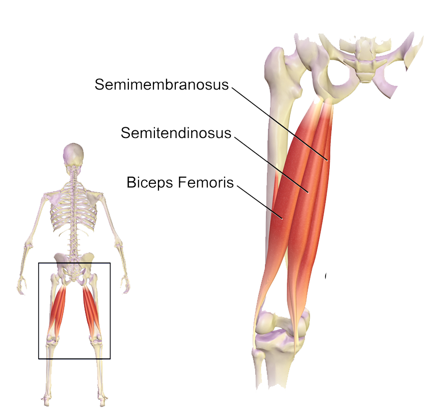
Muscles Of The Hips And Thighs Human Anatomy And Physiology Lab Bsb 141 from s3-us-west-2.amazonaws.com The information contained in anatomy atlases is not a substitute for the medical care and advice of your physician. Serial cross sections anatomy sartorius muscle, profunda femoris (deep femoral) artery and vein, pectineus muscle lliopsoas muscle, rectus. …front and sides of the thigh. Bf sh, lh, biceps femoris short head, long head; They have a lot to do with how your hips move. A condition known as compartment syndrome most commonly affects the divisions of the lower limb, although the upper to see how complete anatomy can transform your learning, at home or in the classroom, try it for free. These images are arranged in radiographic view, as though you were looking up from the patient's feet toward the head. Anatomynote.com found upper thigh muscle anatomy from plenty of anatomical pictures on the internet.
The information contained in anatomy atlases is not a substitute for the medical care and advice of your physician.
The thigh muscles don't just move your legs. Because the hamstrings cross the back of the hip joint on their way to the knee, they help to extend the hip. Muscle and tendon characteristics classic human muscles of the leg and foot classic human anatomy in motion: Pelvic & upper thigh anatomy. There is an obvious constriction which marks the base of the head with the upper portion of the neck, and this also marks a very common site for fractures in the elderly. This muscle includes four heads that originate in different locations but all share the. The muscles of the anterior part of the thigh include the quadriceps group and a few others: Both the thigh and leg are divided into three separate compartments. Serial cross sections anatomy sartorius muscle, profunda femoris (deep femoral) artery and vein, pectineus muscle lliopsoas muscle, rectus. Thus, it is thicker in the upper and lateral part of the thigh, where it receives a fibrous expansion from the glutæus maximus, and where the tensor fasciæ latæ is inserted between its layers; This arrangement gives the hip anatomy a large amount of motion needed for daily activities. 630 anatomical structures of the upper limb (pectoral girdle, shoulder, arm, elbow, forearm, wrist we used the terminologia anatomica to label all the anatomical structures; Anatomically, it is part of the lower limb.